By: Frank Van Campen en Hans Huijbregts
Microscopical observations start with preparations, of course. Largely written out of the historical narrative, the MicroLabs demonstrated the crucial importance of preparing (parts of) insects, plants and trees for minute observation. With regard to flora: only some objects are small and thin enough to be examined directly under a microscope. Most objects must be cut into thinner slices, or sections, to allow sufficient light to pass through them for microscopical examination. In the seventeenth century only razors and pen knives were available for this purpose. And very few references to the staining of seventeenth- century preparations can be found in the literature.
The instruments and techniques used in light microscopy today are mainly based on techniques that were only developed in the nineteenth century. If we compare the present-day techniques with seventeenth-century techniques, the current use of complicated cutting machines (microtomes) and often very complicated staining protocols are particularly striking. In this blog we describe the twentieth-century techniques for cutting sections of plant material.
Hand cutting sections with a ‘Gillette’ razorblade
This procedure can be used for cutting small roots, stems and leaves as long as they are not too hard. During cutting, the plant material is gently pressed under an object slide. This method is mainly used for temporary slides because the cuttings are usually too thick.
-

- 1. Cutting Slices with Gillette Razorblade
-
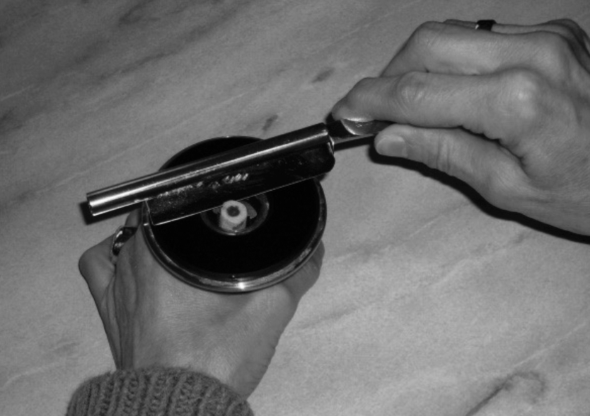
- 2. Cutting slices with a razor and a handmicrotome
Cutting sections with a hand microtome
Plant material is fitted into a supporting material such as elder pitch or a suitable piece of carrot. Then the supporting material is clamped firmly in the microtome and the clamp is raised until the object protrudes from the cutting table. The cutting is carried out using a sharp razor blade that is moved in a continuous sliding motion. The above method can be used for roots, stems and leaves. Depending on the object, a chemical pre-treatment may be required. Cutting thicknesses of 30-50 µm are possible.
-
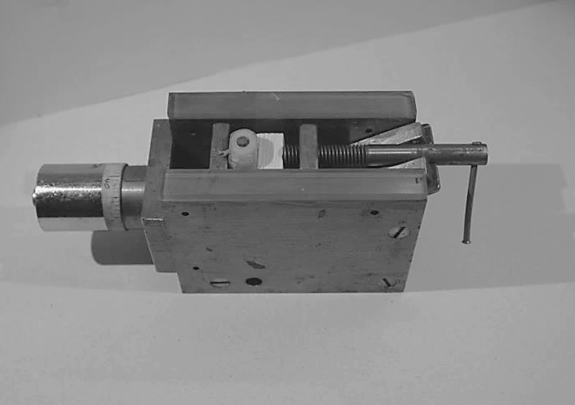
- 3. Lelong microtome, also useful for wood sectioning
-

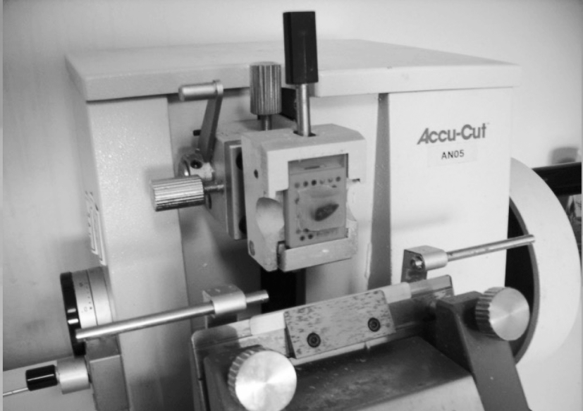
- 4. Cutting paraffine sections on a sliding microtome
Cutting sections of wood
Microscopic examination of wood usually requires special techniques due to the hard and tough properties of wood. Before cutting, small wood cubes of about 10 mm square are boiled in water for several hours to weaken the structure of the wood. These wood blocks are cut in three different planes (transverse, radial and tangential) to make all anatomical structures clearly visible. The cutting is usually performed on a mechanical sliding microtome using a sturdy microtome knife especially suited for wood. The so-called Lelong microtome is a less advanced manually operated microtome that is very suitable for cutting wood (see photo 3). Cutting thicknesses of 30-50 µm. Really fresh soft wood, like willow can be cut without previous treatment.
Cutting sections of wax embedded objects
Some objects such as flower buds, hollow stems and animal tissues can only be cut with additional support from embedding material. This requires a multi-step process with different chemicals. In short, the tissue is first impregnated and then embedded in a suitable material, such as molten paraffin wax, to obtain a stiff block of material. This block is then cut on a sliding or rotary microtome. After cutting the wax is removed from the sections. In this way better and thinner cuts are made than with methods 1-3. The thickness of the cuts varies from 5-10 µm (animal tissue) to 10-20 µm (plants).
-
- 5. Cutting wax embedded preparations
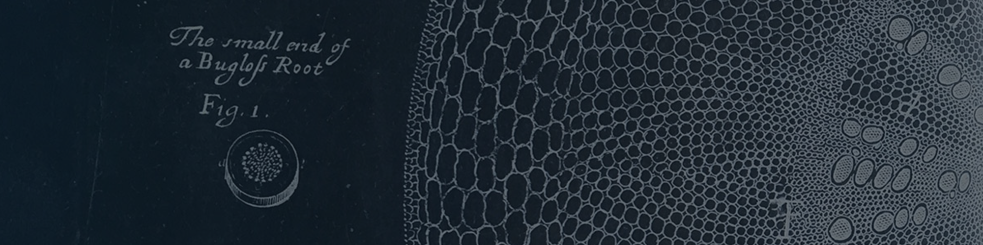
Contrary to these modern techniques, in Microlab 5, we have explored the possible techniques that microscopists in the seventeenth-century may have used to make plant sections. Not having acces to micrtomes or Gillette razors, we wondered how Nehemiah Grew, Antoni van Leeuwenhoek and Marcello Malpighi made slices that were thin enough to yeild good obervations with the micrscope. Something they evidentially managed to do, as their beautiful images of sectioned plants and roots demonstrate. Did they use their own razors? A kitchen knife? Or did they borrow the woodshavings from a carpenter’s workshop? During the MicroLab we tried different approaches to achieve thin sections, as such practical issues were never described. And noticed that reverse engineering from 20th century techniques provided much food for thought.
Are you curious about microscopy? And how things are done now, instead of in the 17th century? More information–and inspiration–may be found on the websites of the Nederlands Genootschap voor Microscopy and Koninklijk Antwerps Genootshap voor Micrografie