This project re-examines the emerging study of the micro-world in the seventeenth century. In traditional accounts of the Scientific Revolution, the microscope is considered the twin-instrument of the telescope. With the latter, Galileo discovered the satellites of Jupiter in 1609, and Christiaan Huygens the ring of Saturn in 1656.
With the microscope, luminaries as Robert Hooke, Marcello Malpighi, Johannes Swammerdam and Antoni van Leeuwenhoek would fundamentally change human understanding of the micro-world. Observations of the microcosmos (insects, bodily fluids and bacteria) – as for example in Hooke’s stunning Micrographia (1665) – seemingly mirrored the spectacular celestial discoveries.
However, we challenge the traditional parallel narratives on the rise of microscopy and astronomy. Besides the nature of the microscope itself, the objects of study differed fundamentally from celestial bodies. The latter could only be seen from a great distance. Insects on the other hand, had to be caught, cut or dissected, and manipulated, under different lighting conditions. This is what we actually tried, hands-on. We literally took a fresh look, with eye-catching results.
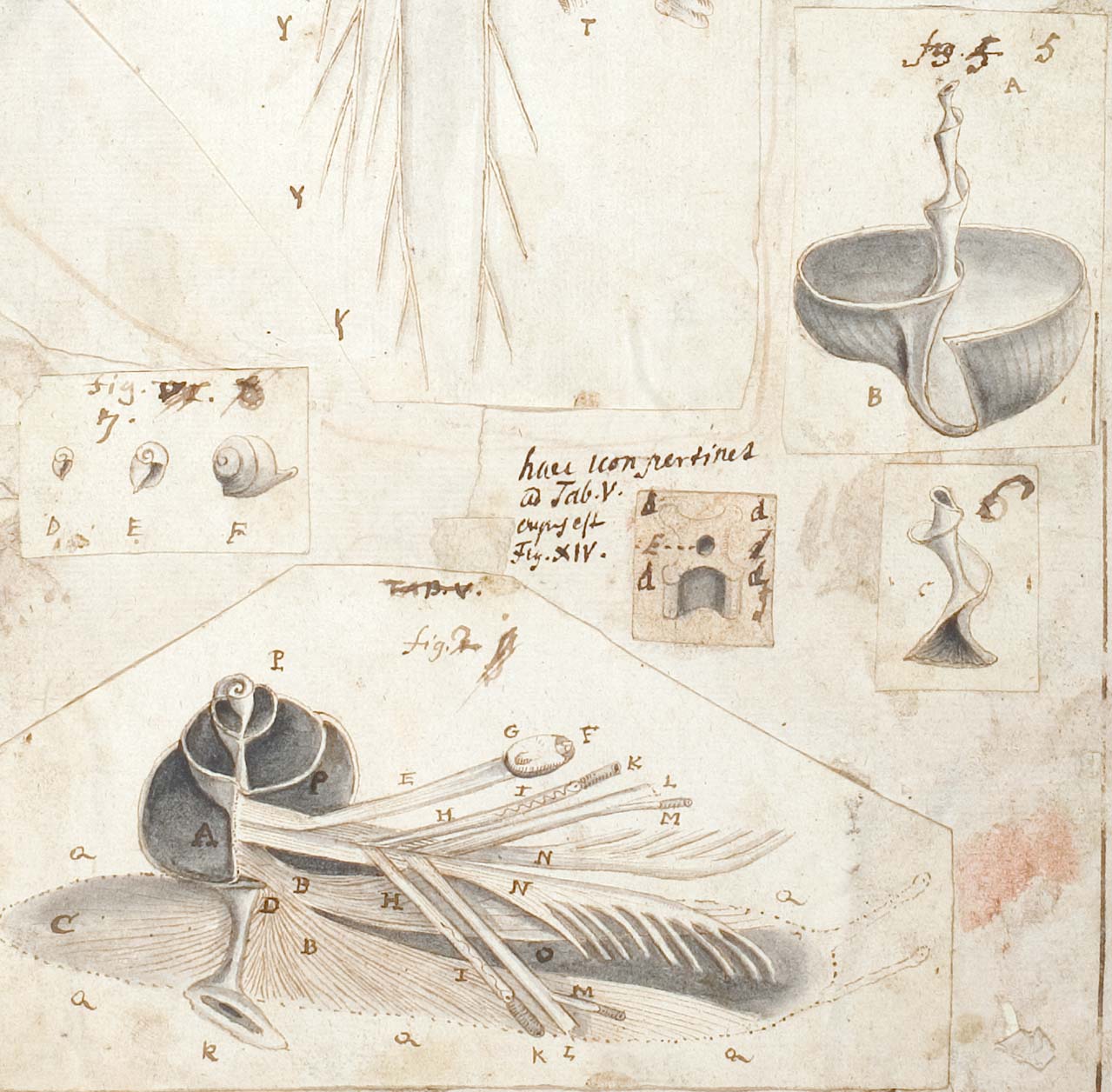
We took the surviving publications, notes and drawings as our point of departure. By using original seventeenth-century microscopes, we tried to reconstruct the original observations. We found out that behind a seemingly snapshot-like image, a range of different techniques and strategies lay hidden.
Much more than the magnification of the instrument, techniques of dissecting, the source of light, and artistic choices were factors at play. What at first glance seems an unequivocal image, is in fact an heavily constructed work of art, based on many dissections and observations. Some details were highlighted – and others left out.