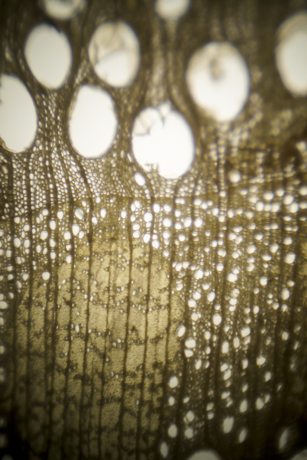
In Microlab 5 the Visualizing the Unknown-team turned its attention to botany. In emulation of the pioneers of plant anatomy – Marcello Malpighi, Robert Hooke, Nehemiah Grew, Antoni van Leeuwenhoek – we tried to reconstruct the preparation and observation techniques of this field of study, largely uncharted territory in the last decades of the 17th century.
In order to create a context for the early microscopical research into botany we discussed analogies and possible influences on the visual language of the botanical images produced by the pioneers mentioned above. Did they look towards anatomical illustrations (human or animal) for inspiration? Did the microscopists influence each other? Was the visual language particular to plant anatomy? In this discussion we were delighted to have Pamela MacKenzie in our midst as a guest. She recently completed her thesis on Nehemiah Grew.
But after these theoretical discussions we quickly got down to the practical handiwork of reconstructing (or reverse engineering) the research practices of the microscopical pioneers. Once again Van Leeuwenhoek’s reports in his letters proved quite informative on this topic. And – as in the earlier Microlabs – practical re-enactment proves to be very useful in filling in the ambiguities left after reading van Leeuwenhoek’s reports.
Our activities focused on producing specimens of sufficient quality to allow useful microscopical observations. That is to say slices of plants thin enough to reveal their structure, either in cross sections or in longitudinal sections. Experiments with a very sharp block plane turned out less successful than we had hoped. Much better results were achieved by using a very sharp old fashioned razorblade. Using the razor to cut very thin slices, or shavings really, of oak, cork, thistle etc. we succeeded in replicating quite closely the sections of plants and tree-bark known
from the illustrations of Van Leeuwenhoek, Grew or Hooke.
Another point of interest for the research team was the influence of lighting the specimen on the image seen through the microscope. Lighting the specimen from behind produces a very different image than lighting it from above. As not all microscopes of the era allowed lighting from behind, the images offered in the work of the early microscopists tell us something about the instruments they used.
