About

This project critically examines the emerging study of the micro-world in the seventeenth century, with a focus on the manner in which images were created and came to be used. In most accounts of the rise of modern science, the microscope is considered the twin-instrument of the telescope. With the latter, Galileo discovered the satellites of Jupiter in 1609, and Christiaan Huygens the ring of Saturn in 1656.

With the former, the microscope, luminaries as Robert Hooke, Marcello Malpighi, Johannes Swammerdam and Antoni van Leeuwenhoek would fundamentally change human understanding of the micro-world. Observations of the microcosmos – insects, bodily fluids and bacteria –, as for example in Hooke’s stunning Micrographia (1665), seemingly mirrored the spectacular celestial discoveries.

However, we challenge the traditional parallel narratives on the rise of microscopy and astronomy. We wonder what actually counted as a microscope, could magnifying glasses also count? What were fundamental differences between the microscope, magnifying glasses and single-lens microscopes? And when considering the latter – as used by Swammerdam and Leeuwenhoek _ , what are the visual strategies used and how do they change throughout the sixteenth and seventeenth centuries? How important was the scale of magnification?

Besides the nature of the instrument itself, the objects of study differed fundamentally from celestial bodies. The latter could only be seen from a great distance. Insects on the other hand, had to be caught, cut or dissected, and manipulated, under different lighting conditions. The moon, sun and planets had been depicted since antiquity. So were all kinds of animals. But a visual idiom for understanding and representing the new micro-world had to start from scratch. Visualizing the Unknown explores these issues.

Background image: Head of a bee, as seen through a Van Musschenbroek microscope, magnification 30x. Cat. nr. V07512. Rijksmuseum Boerhaave © Wim van Egmond for Visualizing the Unknown

The Project

Visualizing the Unknown takes a fresh look. We will start watching and recording through various seventeenth-century lenses – including the iconic ones by Leeuwenhoek. What can we see? What could have been seen? What were the challenges, problems and pitfalls? Did preconceived notions influence the observations? And – crucially – how were observations made by single person, translated into an image that could convince others? Our multidisciplinary team brings together innovative ideas and practices, coming from various angles. Embedded in the Huygens Institute, the Bibliotheca Hertziana – Max Planck Institute for Art History and Rijksmuseum Boerhaave, we can build on extensive knowledge of the history of science, art history and the materiality of instruments. Importantly, some of our team-members are trained at art school.

The six-year project will, stage by stage, explore the entire trajectory of microscopical observation to publication and impact. It explores previously unchartered facets in the making of knowledge. Our subject and time-span coincides with the emergence of scientific societies, of scholarly journals, and the practice of peer-review. As such, Visualizing the Unknown is of great relevance for our understanding present-day scientific communication, including the use of images.

Although some of the pioneers of microscopy have become famous, many others were destined for oblivion. We will not write an history of exceptional figures and their discoveries. Instead, we will reconstruct local and international networks, giving special attention to the lively exchange of knowledge, objects and publications between England, the Dutch Republic and Italy.

Then, as now, in science the context of discovery and the power of proof and persuasion were key. Discoveries and the establishment of facts are not self-evident, but the result of complex heuristic processes and negotiations. Were the facets of a bee’s eye circular or hexagonal? Did silkworms have a brain and, if so, how to depict it? Did blood-cells consist of globules? And there always were issues of priority disputes, openness and secrecy. Whom was to be trusted? How was authority constructed?

Our project will also contribute to two major exhibitions at the Rijksmuseum and Rijksmuseum Boerhaave. Visualing the Unknown will result in a documentary, a program of education for primary schools, and activities commemorating Leeuwenhoek in 2023. Our output will include publications – articles, a dissertation and various books – and when the time has come, open-access to a database with the spectacular photos and videos our award-wining photographer took through original Leeuwenhoek, Van Musschenbroek and Campani microscopes.

The Team of Visualizing the Unknown keeps you updated on ongoing research with a Blog, by regulary updating our Publications and Events. And by posting on social media such as Twitter and Instagram. 

Van Leeuwenhoekmicroscoop, 1670-1710. Silver and Glass. V07019. Rijksmuseum Boerhaave. Stack Photographed by Wim van Egmond
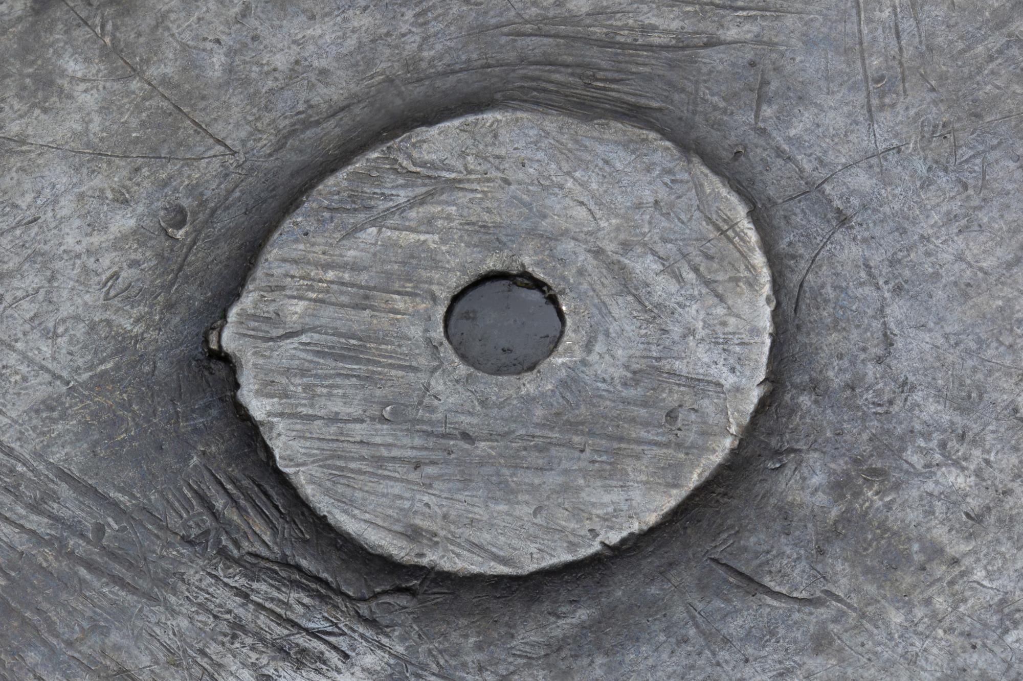
Mounted lens (close up) of Antoni van Leeuwenhoek microscope, magnification 68x. Cat. nr. V30337. Rijksmuseum Boerhaave. Stacked macro photograph © Wim van Egmond for Visualizing the Unknown

Method

The project has started  with minute scrutiny of original notes, drawings and publications and – in a way that has never been done before – preserved historical instruments or reconstructions specially made for our research in our MicroLabs. We  have tried  to approach what Malpighi, Swammerdam and Leeuwenhoek saw and which problems they could have encountered by redoing their observations. We did this in a lab-setting, provided by Rijksmuseum Boerhaave, making use of the institution’s impressive collection of seventeenth-century microscopes as well as the latest digital techniques. With our approach we elaborate on the method of re-enacting proposed by for example Pamela Smith – member of our advisory board – as well as  try to follow the trail back from (still existing) accounts and original drawings, now scattered throughout archives and libraries in Europe, to understand better how observations with the microscope were translated into text and image.

The project forms a continuation of earlier research by some of the team members on sources by Swammerdam and Leeuwenhoek, a Cambridge-based project on the visual culture of the early Royal Society (“Making Visible”), and research on early modern optics. During a pilot-project in 2019, some of Leeuwenhoek’s original specimen -now in the Archives of the Royal Society- were brought to Rijksmuseum Boerhaave. The specimen were viewed through his original microscope, leading to spectacular results that were documented in photographs and on video.

Research Process

A silkworm’s trachea, as seen through a Campani microscope, magnification 15-20x. Cat. nr. V28600. Rijksmuseum Boerhaave © Wim van Egmond for Visualizing the Unknown

Our official kick-off meeting took place on 1 October 2021. The project consists of  subsequent stages. In the first phase, the entire Team – including our PhD-student – reconstructed the observations done with the microscope by our protagonists. Starting in 2024, we will research how drawings were constructed, interpreted, reviewed communicated through the European world of learning. With a strongly visual approach we will also use the images made in the first phase of the project to compare them with the historical material. In the third phase, a post-doc will give special attention to various visual techniques, such as ad vivum-representations, time-lapsing, nature-prints, models and the inclusion of insect-parts in paintings. We contend that not only the publication of books and journals formed part of the trajectory of knowledge-production and communication, but also included cabinets of curiosity and works of art.

Sharing knowledge and experience forms the basis of our approach. In the spirit of the early modern Republic of Letters, we will take an international perspective. Visualizing the Unknowns crosses disciplinary as well as geographical boundaries. We will not confine ourselves to study the work of well-known microscopists in isolation. We choose for a comparative approach, including lesser-known figures (at least for their work as microscopists), such as Athanasius Kircher, Nicolaes Hartsoeker and Filippo Buonanni.

The project-team organizes and contribute to international workshops and conferences. The project will be concluded by a richly illustrated monograph, scheduled for 2027.

Key Figures

Prince Federico Cesi, Founder Accademia dei Lincei

Prince Federico Cesi

1595-1630

Prince Federico Ceci founded the Accademia dei Lincei in Rome in 1603. The academy aimed at understanding nature and her secrets through close observation and experiment. The Lincei started using the word ‘microscope’ for compound magnifying instruments in a play on the word ’telescope’. Members of the Lincei were also the first to explicitly make use of microscopes for the production of images that represented the anatomy of the bee magnified.

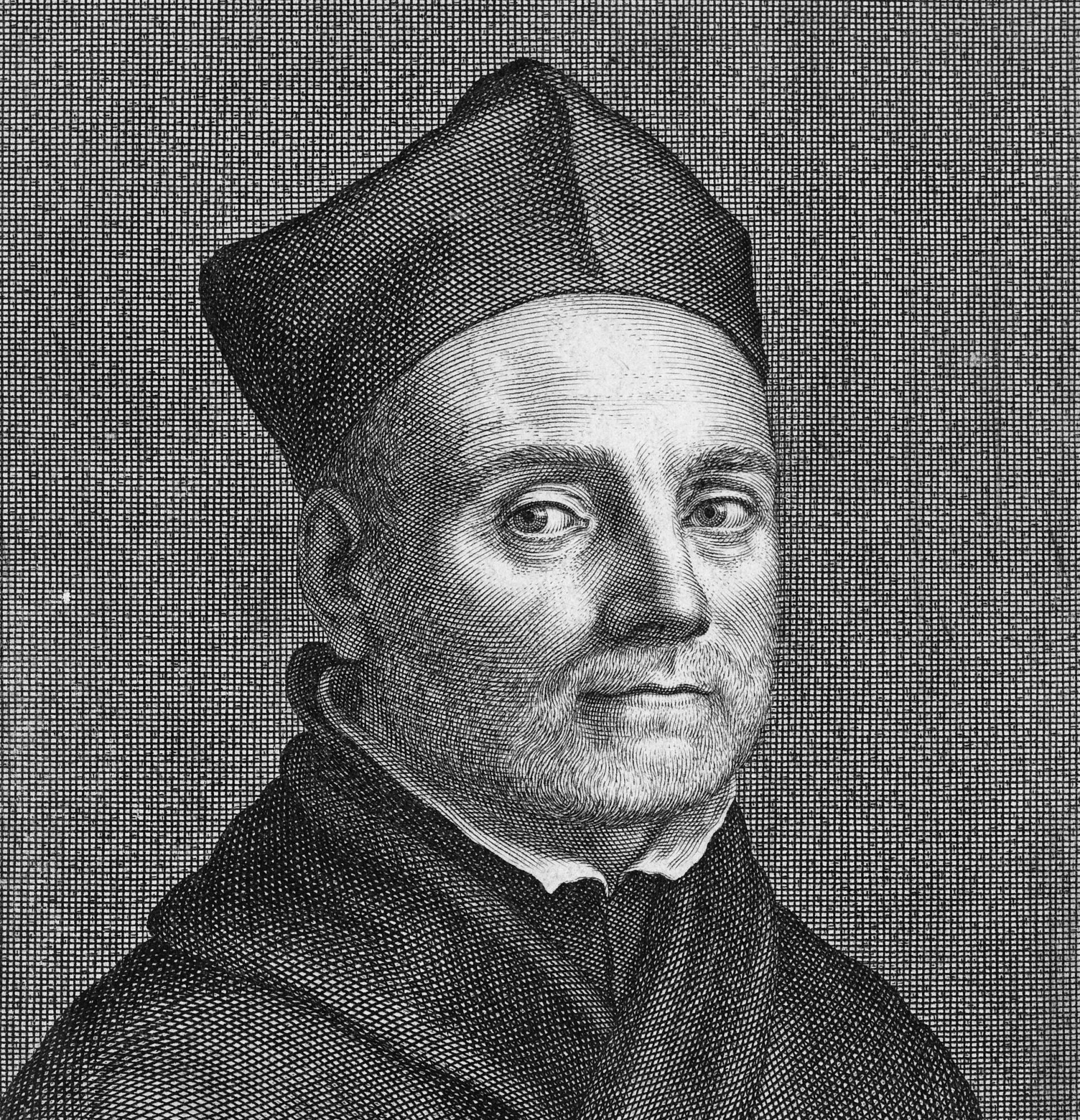
Athanasius Kircher, Father Kircher, Jesuit

Athanasius Kircher

1602-1680

Trying to better understand the bubonic plague after living through the devastating Italian plague year of 1656, the Jesuit Priest Athanasius Kircher turned to the microscope. Kircher started to examine the blood of plague victims under his magnifying instrument (which he called smicroscopus) finding  it was filled with thousands of tiny little wiggling worms. Studying a disease with scientific instruments such as microscopes may seem intuitive now, but Kircher was probably one of the first—if not the first—to do so.

Robert Hooke, Eye of the Fly, Eye of Fly, Hooke, Micrographia

Robert Hooke

1635-1703

Robert Hooke could do many things well; he was a great mathematician, physicist, and draughtsman. He was appointed curator of experiments for the Royal Society in 1662 and often cooperated with Robert Boyle. In 1665 Hooke published his Micrographia, wherein readers find exceptionally detailed copperplate engravings of common objects and creatures put under the microscope. The book was an enormous success; inspiring microscopic research in years to come. And revealing that drawing was an essential skill for 17th century microscopists to record and disseminate their observations.

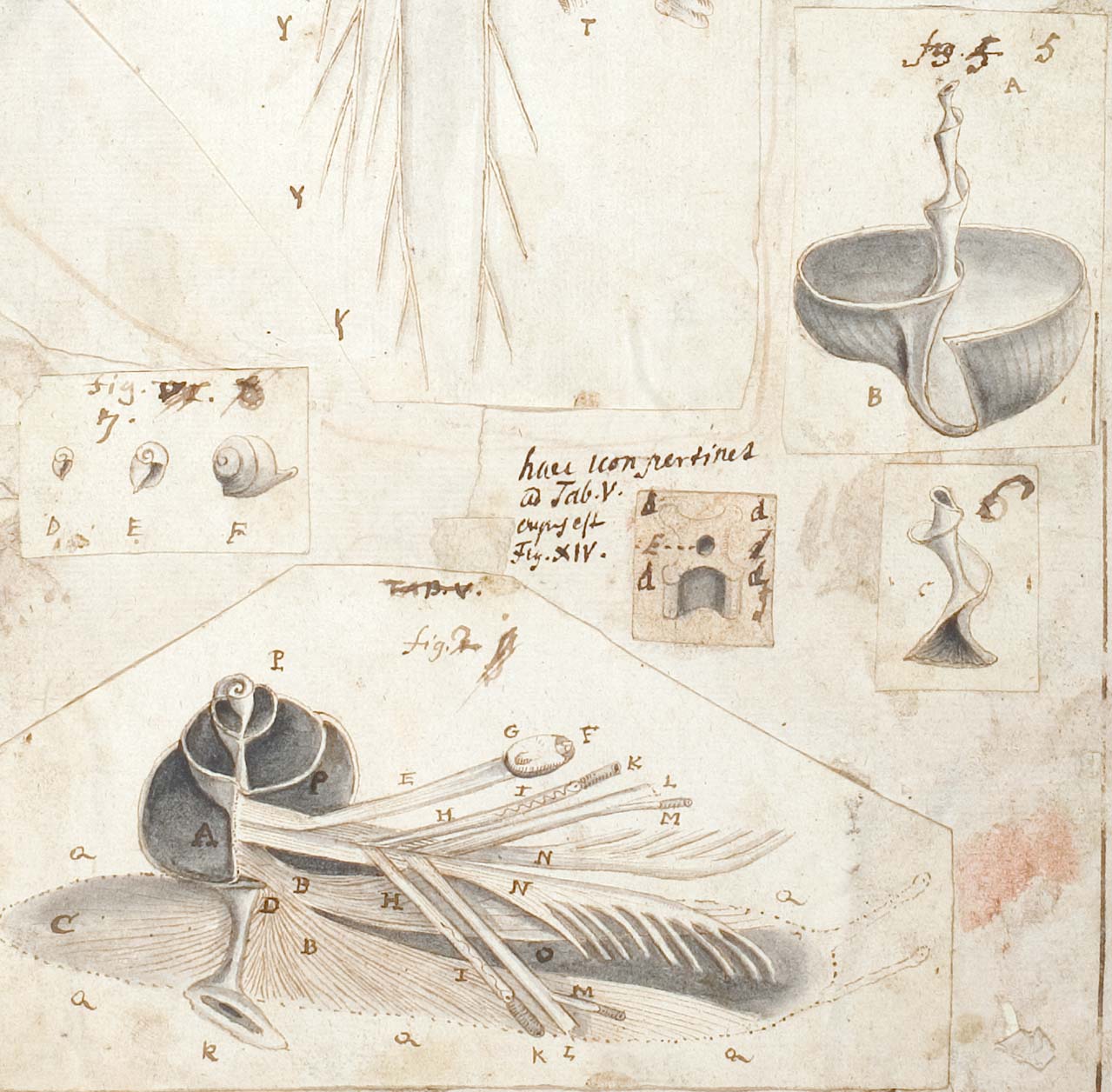
Johannes Swammerdam, The anatomy of a snail in Icones operis Bibliae naturae c. 1678. Drawing, detail from fol. 7r. BPL 126 B. Leiden University Library

Johannes Swammerdam

1637-1680

Johannes Swammerdam was one of the first to study the inner anatomy of insects with the use of single lens microscopes. Using tiny scissors and pincers, he revealed insects and other creepy crawlies had a very complex inner anatomy. By unveiling this, Swammerdam disproved the Aristotelian theory that insects had no inner anatomy at all. And generated spontaneously from putrefying matter. Indicating the ‘Finger of God’ in the anatomy of a louse. Importantly, Swammerdam not only managed to see insect anatomy with his microscope, he also represented his observations in intricate, well thought-through drawings.

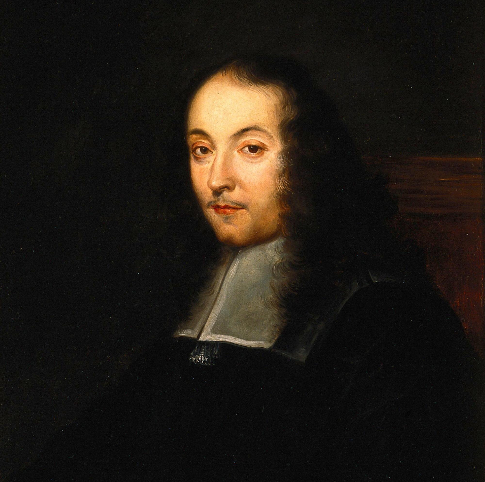
Anonymous artist, portrait of Marcello Malpighi, oil painitng, Wellcome Collection London, refference: 45748i

Marcello Malpighi

1628-1694

Using the microscope to investigate the structures of human tissue, insects, amphibians and plants, Marcello Malpighi was a pioneer of microscopy. Malpighi dissected silkworms for an assignment of the Royal Society, managing to uncover and draw its delicate anatomy. The drawings were made into copperplate engravings for Malpighi’s subsequent publication, titled De Bombyce (1669). This publication inspired Swammerdam, who then also began to investigate the structure of silkworms with his microscope. Swammerdam wanted to figure our Malpighi’s methods, and see if he could confirm the Italian’s observations. In a similar turn of events, Malpighi investigated plant anatomy around the same time as Nehemiah Grew, allowing  for comparison and confirmation of observations made with the microscope.

Portrait Engraving Francesco Redi, C. 1670, Wellcome Collection

Francesco Redi

1626-1697

The Tuscan poet and physician Francesco Redi was the first to challenge the theory of spontaneous generation by demonstrating that maggots found in putrefying meat resulted from eggs laid by flies. In his publication on this topic, Esperienze Intorno alla generazione degl’insetti (1668), Redi also included images of ticks, lice and other parasites. These images were made with the help a microscope. Later he would publish a volume on parasites alone, including images of body parts of dissected parasites. For his work Redi is considered a founder of experimental biology and parasitology.

Jan Verkolke, Antoni Van Leeuwenhoek, 1680-1686, Rijksmuseum Amsterdam

Antoni van Leeuwenhoek

1632-1723

Starting late in his life with microscopy—he was already in his 40s—Antoni van Leeuwenhoek became virtually obsessed with the microworld. He engineered his own type of single lens microscope, while perfecting the lenses he used. With them, Van Leeuwenhoek enjoyed studying all sorts of things. Resulting in new discoveries, such as: bacteria, single-cell organisms, spermatozoids, and red blood cells. Van Leeuwenhoek frequently shared his microscopic observations with the Royal Society. Leading Robert Hooke back into conducting research with the microscope, in an effort to confirm Leeuwenhoek’s observations and to reverse engineer the Dutchman’s methods.

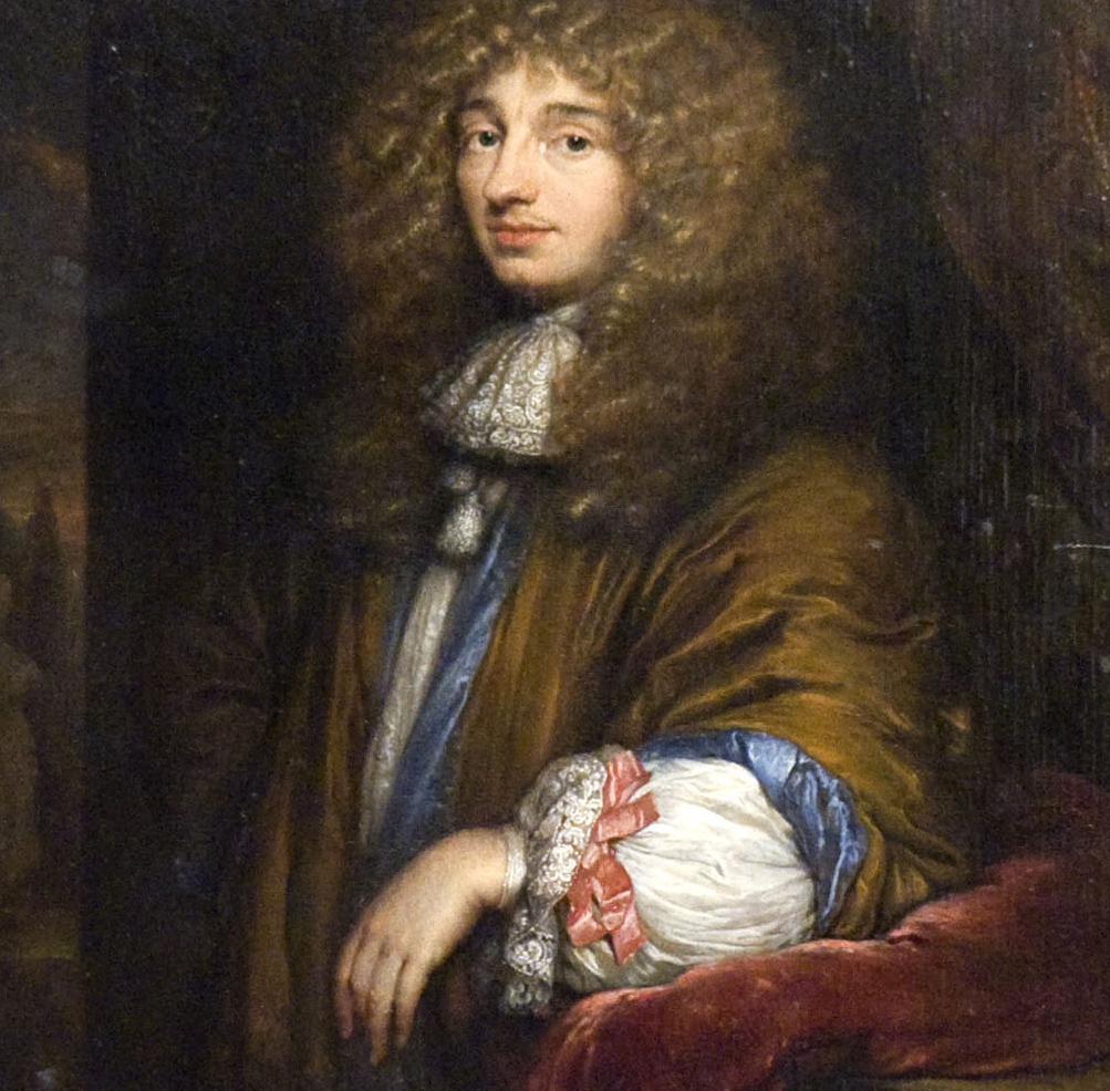
Christiaan Huygens, Huygens

Christiaan Huygens

1629-1695

Christiaan Huygens, member of the Royal Society and the Académie des Science, is probably most famous for his improvement of and work with the telescope. But he also used microscopes during his long scientific career. He checked the observations of Van Leeuwenhoek, whose work prompted him to start using single lens microscopes instead of compound microscopes. As well as to draw what are now the earliest  representations of spermatoziods. Huygens was acquainted with Nicolaas Hartsoeker and introduced him to French scientists such as Nicolas Malebrache and Giovanni Gassendi while in Paris.

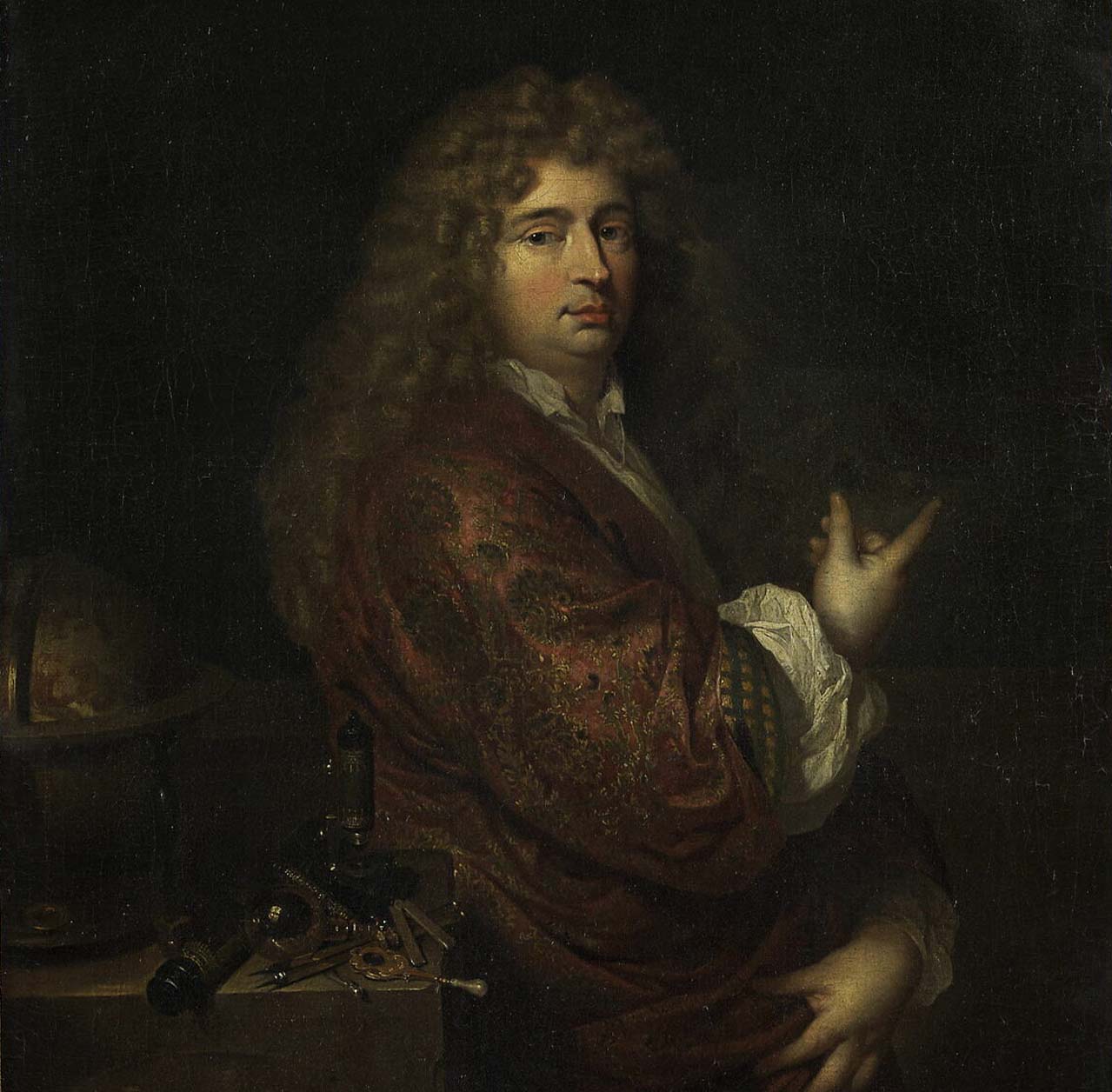
Nicolaas-Hartsoeker; harsoeker;microscopists

Nicolaes Hartsoeker

1656-1725

Known for his competition and rivalry with Antoni van Leeuwenhoek, Harstoeker is an interesting figure in himself. He became interested in optics in his early twenties, and started making money as a lens grinder when he lived in Rotterdam. Hartsoeker met Christiaan Huygens in 1677,  teaching him how to make single lens microscopes. A year later Hartsoeker would accompany Huygens to Paris. After a brief return to Holland, Hartsoeker would go to Paris as a scientific instrument maker. At the end of the 17th century Hartsoeker was famous and elected a member of the Académie des Sciences in 1699 and the Königlich-Preußische Akademie der Wissenschaften in 1704.

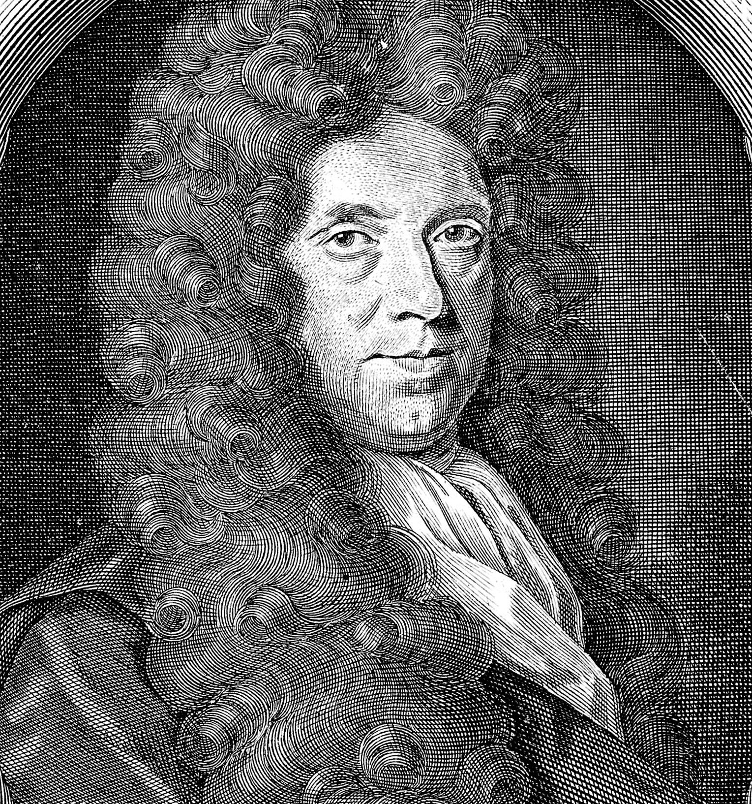
Nehemiah Grew, Grew

Nehemiah Grew

1641-1712

The Royal Society received two manuscripts on the same subject in the same year: One by Nehemiah Grew the other by Marcello Malpighi. Both naturalists had used microscopes to study the textures and layers of plant anatomy. Though this could have led to rivalry, they saw the use of the other’s work, as now they were able to confirm each other’s observations. Grew’s publication, The Anatomy of Plants begun (1682) included 82 beautiful and elaborate copperplate illustrations. In addition, Grew served as secretary of the Royal Society along with Robert Hooke after the former secretary, Henry Oldenburg, passed. Grew edited the Philosophical Transactions in 1678–1679.

Colophon, Copyrights and Disclaimer

This is the colofon and disclaimer of Visualizing the Unknown. This disclaimer is relevant when you use our website. Visualizing the Unknown makes every effort to provide timely and accurate information on this website. Nevertheless, there may be errors and/or information that is incomplete and/or contains inaccuracies. Visualizing the Unknown does not assume any liability for the relevance, accuracy, completeness or quality of the information provided. Visualizing the Unknown rejects any direct or indirect damages incurred from the use of our website, and shall not be liable for damage of a tangible or intangible nature caused directly or indirectly through the use or failure to use the information offered, and/or through the use of faulty or incomplete information. Visualizing the Unknown reserves the right to modify, supplement, or delete any or all of the information offered on its internet site, and/or to temporarily or permanently cease publication without prior and separate notification.

Copyrights:

This site contains copyrighted materials. All texts were written by our team. All microphotographs were made by Wim van Egmond. It is not allowed to copy or spread texts and/or images from this website without expressed written permission.

Third Party Rights:

Visualizing the Unknown has done its best to contact and ask permission of owners for the publication of materials and data shown on this website. Please contact us when you believe that certain shown materials breach your copyrights. Also contact us when certain published data or materials are incomplete, or factually incorrect, or given unclear or incomplete reasons for inclusion on this website. We will contact you as soon as possible. When a comment or complaint is well-founded, we will remove or correct materials or contact you to see how we can accommodate your report.

Legal Structure: 

Visualizing the Unknown is a NWO funded non-profit research project. We are partnered with: the Huygens Institute (KNAW-Royal Netherlands Academy of Arts and Science, Amsterdam), the Bibliotheca Hertziana–Max Planck Institute for Art History (Rome) and Rijksmuseum Boerhaave (Leiden). Additional partners are Leiden University, the Royal Society (London) and the Rijksmuseum (Amsterdam).

Colophon:

Copyright: © 2021-2027 Visualizing the Unknown

Photography: Microphotography/microlabs © Wim van Egmond for Visualizing the Unknown, Team Portraits © Fred Ernst, Historic microscopes © Tom Haartsen for Rijksmuseum Boerhaave

Authors: Prof. Dr. Eric Jorink, Dr. Sietske Fransen, drs. Tiemen Cocquyt, Dr. Mieneke te Hennepe, Dr. Tim Huisman, Geertje Dekkers, Ellen Pater, Larissa van Vianen

Graphic Design: Ellen Pater

Web Development and Built: Madpack & Brendly

Made Possible by: Huygens Institute, Amsterdam; Biblioteca Hertziana – Max Planck Institut für Kunstgeschichte, Rome; Rijksmuseum Boerhaave Leiden